

Dr Rocío Lazo, Specialist in Paediatric Dentistry at Cayetano Heredia University of Peru, discusses the role of bioactive restorative materials and techniques and their role in minimally invasive dentistry
I have been a paediatric dentist since 2002 and, for the last 12 years, I have been involved in research about minimally invasive dentistry and the use of bioactive biomaterials, which are proving to be something of a revolution in dentistry.
What is minimally invasive dentistry?
Minimally invasive dentistry (MID) is the application of a systematic respect for the original tissue. The aim for dentists who use this approach is to focus on removing as little as possible when it comes to the tooth structure while at the same time getting rid of the problem and improving the patient’s general oral health. For treating carious lesions or Molar Incisor Hypomineralisation (MIH), MID offers three different treatment options:
- When treating enamel carious lesions (pit and fissures or interproximal caries) it is not always necessary to do a restorative treatment. MID suggests a non-restorative cavity control treatment (NRCC). This means not removing the lesion and applying materials to attempt to remineralise them. NRCC could be an option to manage carious lesions on occlusal or proximal surfaces of primary and permanent teeth.
- In the case of enamel caries that require restorative cavity control treatment, again there is no need to remove the lesion and instead only apply a restorative material. However, these restorative materials need to have special characteristics as found in bioactive restorative materials.
- When caries are in dentine, it is preferable to do selective caries removal, i.e., only removing the infected dentine and sealing the cavity with a remineralised restorative material.
In the case of selective caries removal, infected dentine in the outer dentine caries layer is bacterially contaminated, irreversibly denatured, and impossible of being remineralised. The inner layer is the affected dentine that is biofriendly tissue because it is not infected by bacteria, partially demineralised and the collagen in this zone is intact and physiologically remineralisable.
Selective caries removal in deep caries lesions seem advantageous in comparison to complete caries removal because it helps to preserve the vital pulp. MID suggests selective caries removal in the management of deep carious lesions and indirect or direct pulp capping treatment with bioactive materials in primary teeth. Currently the European Society of Endodontology suggests selective caries removal in the management of deep carious lesions to reduce unnecessary root canal treatment in vital pulp and suggests the indirect or direct pulp capping with bioactive biomaterials (bioactive glass).
Update on MID
Today we know that fluoride varnish alone does not work well in deep white spots carious lesions. Firstly, it only penetrates the surface zone and it is necessary to penetrate into the body of the lesion for the remineralisation of white spots. Secondly, fluoride needs calcium and phosphate to form fluorapatite to combat tooth decay and these zone have less counts of minerals.
What about fluoride in dentine carious lesions? MID suggests only selective caries removal of the infected dentine and not removal of the affected dentine because it is rich in collagen and will be remineralisable. This is important because it is rich in collagen fibres but is demineralised, and fluoride has limited action in re-mineralising affected dentine.
MID needs materials that have the power to penetrate into the body of the lesion in white spots and work to remineralises tissues without minerals.
The problems of MIH
There are many problems associated with MIH which is a particular challenge for paediatric dentistry. In MIH there is a lack of calcium phosphate making it impossible to mineralise with fluoride because fluoride needs calcium and phosphate to form fluorapatite. For example, the application of fluoride varnish (Duraphat) on MIH lesions showed no significant changes in both mean levels of fluorescence and area of lesion over time. One reason may be due to the architectural organisation and protein/mineral content of the affected enamel, which makes it difficult for any attempt of fluoride incorporation to succeed/occur.
The lack of calcium and phosphate in the tissues make teeth easily break down. With MIH the tooth surface is very porous so biofilm is likely to form quickly causing fast development of caries. Another problem in MIH is hypersensitivity which can be extremely difficult to control even with local anaesthesia. The adhesive in teeth affected with MIH is the worst because the protein/mineral content makes an etching pattern type III. The prevalence of MIH is growing around the world and is becoming a major challenge, something that fluoride alone cannot treat.
Materials used in today’s MID
Silver diamine fluoride (SDF) and bioactive biomaterials are ideal materials for MID.
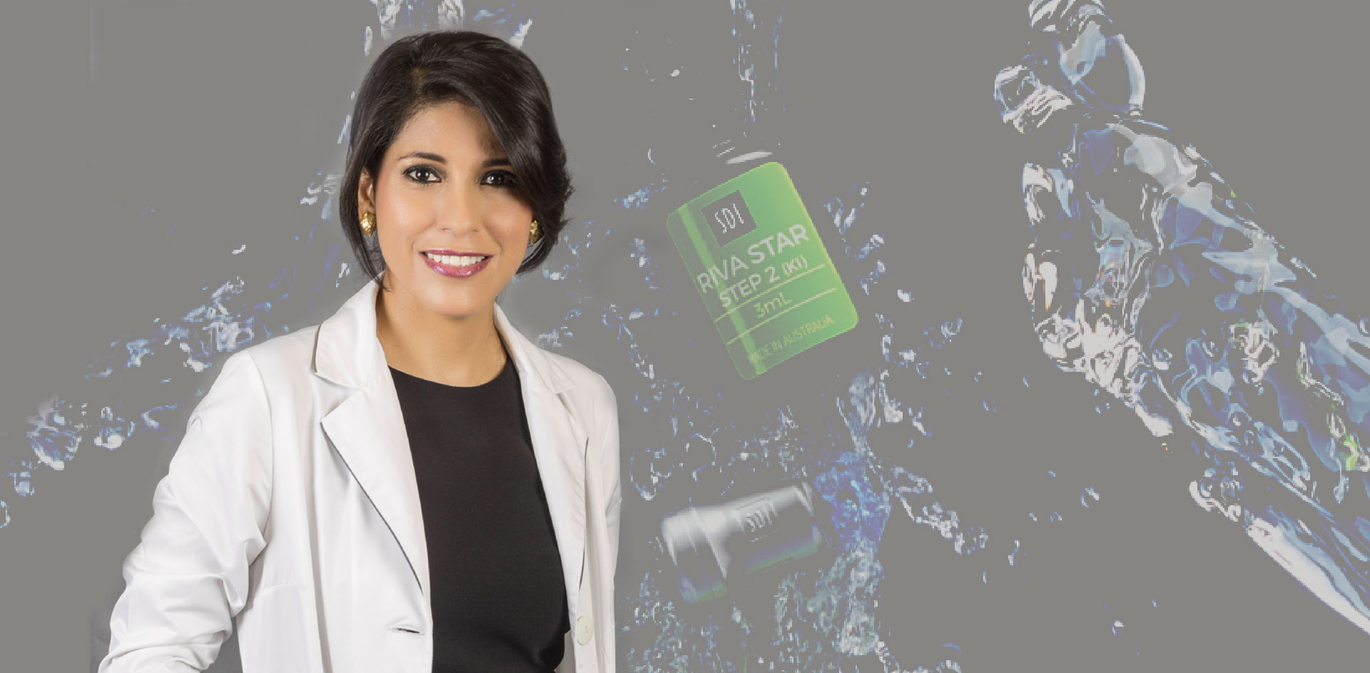
SDF works perfectly to control hypersensitivity, especially in MIH cases. I use Riva Star (SDI Ltd.) to treat hypersensitivity as well for non-restorative cavity control1 treatments where I get great results. A well-known problem with SDF is that it causes staining of the teeth. The reason I like Riva Star and Riva Star Aqua (SDI Ltd.) is because of the patented use of KI which is applied immediately after SDF to minimise staining. Other problems with SDF treatment include the need to apply a gingival barrier to avoid the risk of damage to soft tissue plus the noticeable smell of the ammonium carrier solution. Riva Star Aqua is water-based, meaning there is no risk of tissue damage so there is no need for a gingival barrier, and there is no unpleasant smell.
The SMART technique
The SMART (silver modified atraumatic restorative treatment) is where the carious lesion is first treated by selective removal (only removing the infected dentine) then the affected dentine is treated with SDF and the cavity restored with a conventional or high viscosity glass ionomer modified strontium, effectively arresting caries without removing additional tooth structure. SDF acts as an antimicrobial agent to ensure the removal of all bacteria from the affected dentine to prevent further decay. The GI modified strontium adheres to the tooth structure to seal the cavity, releases fluoride to assist with remineralisation of the tooth and may also work to reduce biofilm formation and recurrent caries.
What is biomimetic dentistry?
Biomimetic dentistry means to copy or mimic the strength, function and aesthetics of natural teeth. Using bioactive biomaterials, it is possible to mimic biomineralisation of enamel, dentine, cementum and bone which is the process of the deposition of apatite minerals calcium and phosphate within the organic matrix. The benefit of biomimetic dentistry is that it preserves the ideal properties of natural teeth using minimal invasive preparation to conserve the enamel and dentine for the best treatment results.
Bioactive restorative materials
Glass ionomer modified strontium is a bioactive material that promotes tooth biomineralisation. Strontium is a natural element present in the body and diet and has the ability to influence the structural property of apatite. Strontium plays an important role on the interaction to hydroxyapatite, forming strontium-apatite.
Strontium and calcium are present in Group 2 of the periodic table. Strontium has an atomic radius slightly larger than calcium and readily substitutes for calcium in minerals. Strontium is known to be able to replace calcium in bone, enamel and dentine.
Strontium-apatite provides an enhanced acid resistance. Studies2 show that the formation of a calcium-strontium apatite complex at the apatite crystal surface retards the acid dissolution. It seems that the strontium concentration interferes directly on the enamel remineralisation process, since when in higher concentrations, rapid remineralisation was observed.
Synergistic effects of fluoride and strontium
Synthesised strontium fluorapatite has been reported3 to demonstrate higher solubility than hydroxyapatite, and the solubility increases with greater strontium substitution. The combined effect of Sr+2 and F– provides more protection to the dental enamel against Streptococcus mutans as compared to their individual effect.
Product choices
Today’s bioactive biomaterials are essential to successful minimally invasive treatments and the product range I use exclusively is SDI Riva, in particular Riva Light Cure HV. Riva Light Cure HV is a GI reinforced resin with fluoride and strontium ions to enhance the remineralisation process and make hydroxyapatite crystals more resistant to dissolution in acid, preventing demineralisation and discontinuing caries progression. Riva Light Cure HV is radiopaque with high compressive strength and can be used for a wide range of restorative options. These characteristics make Riva Light Cure HV an ideal restorative material in MIH and deep carious lesions.
Riva Light Cure HV is also ideal for the Sandwich Technique – a sandwich of GI cement, dental adhesive and composite resin, such as new Luna 2 (SDI Ltd.). The GI is placed as a liner or base, followed by placement of a resin composite to provide greatly improved aesthetic restorative outcomes.
SDI was one of the first companies to research and develop bioactive restorative materials. Riva Star, Riva Star Aqua and the range of Riva glass ionomers are perfect for minimally invasive dentistry and in my experience, SDI’s restorative products always deliver excellent results.
References
- Riva Star is not licenced for caries control in the UK.
- “Novel fluoride and strontium-containing bioactive glasses for dental varnishes-design and bioactivity in Tris buffer solution”, Thaer Jaber Al-Khafajia,b,⁎, Ferranti Wonga, Padhraig S. Fleminga, Natalia Karpukhinaa, Robert Hilla, Journal of Non-Crystalline Solids.
- “Influence of Strontium on the Physical, Mechanical and In-Vitro Bioactivity of Glass Ionomer Cements”, Yiyu Li1*, 1Department of General Dentistry, Peking University, Beijing, 100081
About the author
 Dr Rocío Lazo
Specialist Paediatric Dentist (since 2002)
Professor – Postdoctoral Pediatric Dentistry Program (20 years experience) at Cayetano Heredia University and Científica del Sur University.
Special Smiles Clinical Director by Special Olympics Inc. Internship in Pediatric Dentistry Program University of California San Francisco. 03 Awards Best Research about Medical and Dentistry Science Fundación Instituto Hipolito Unanue-Perú. International Speaker on Biomimetic and Minimally Invasive Dentistry. Invited Lecture THE ROYAL ACADEMY OF MEDICINE AND RELATED SCIENCES OF GALICIA-SPAIN. Publication Co-Author 3 Pediatric Dentistry books and Papers (SDF, XYLITOL, BIOACTIVE MATERIALS)